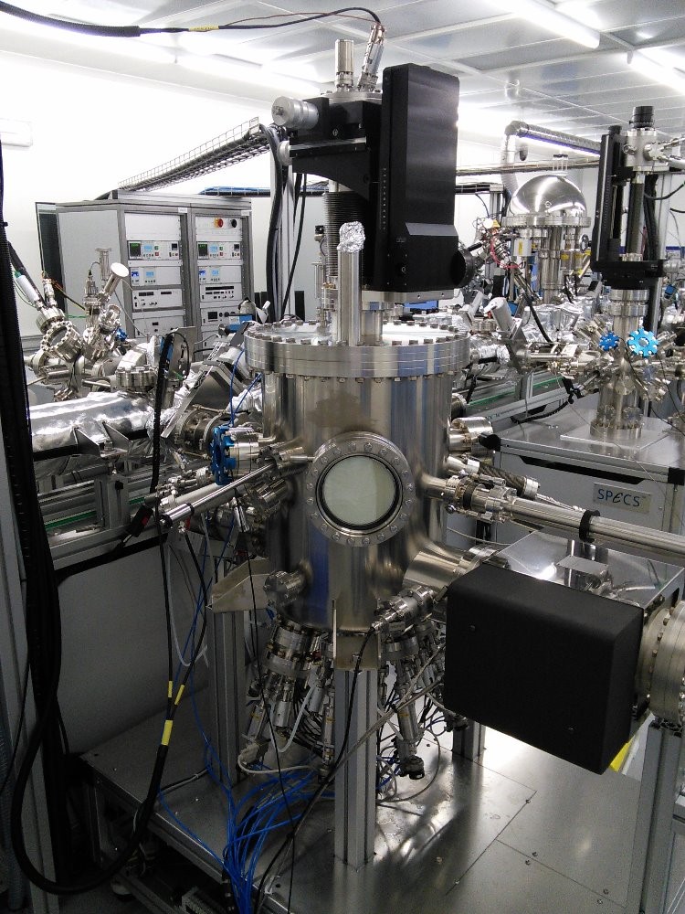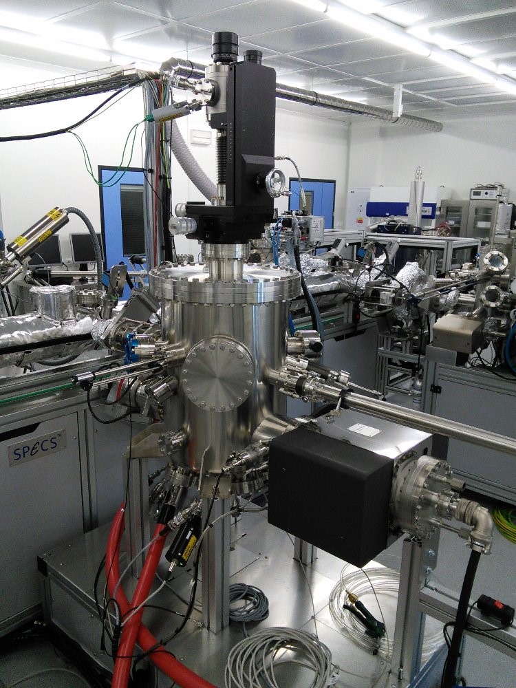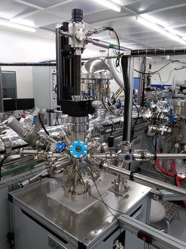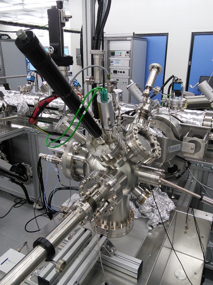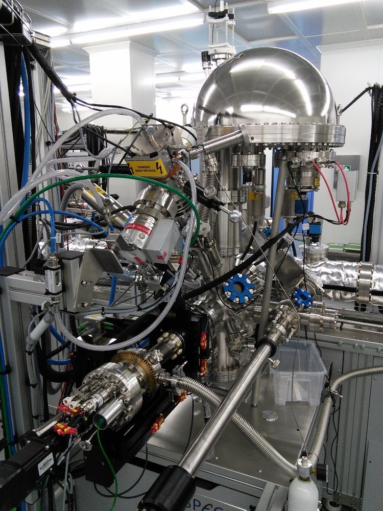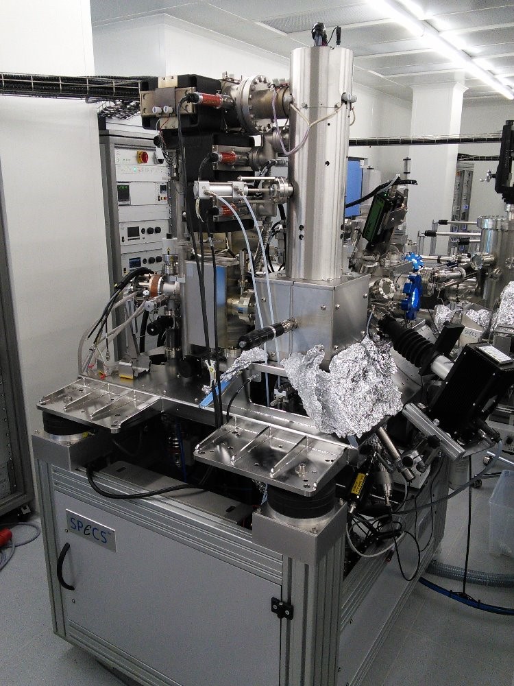
Ultra High Vacuum Preparation and Analytical System (UHV-CLUSTER)
Guarantor
Josef Polčák & Viktor Danchuk
Description
The complex UHV system combines preparation and in-situ analysis by several complementary methods for the characterisation of surfaces and thin films. To maintain a high bulk and surface purity during the sample preparation and subsequent analysis, the whole apparatus is kept under ultra-high vacuum conditions (UHV, base pressure below 2×10⁻⁸ Pa) using an appropriate pumping system. The system comprises 8 individual UHV chambers connected by a linear transfer system allowing sample transfer under UHV conditions and storing up to 15 samples. The individual units are designed to allow users to work simultaneously. The configuration of the apparatus and the selection of analytical methods allows for studying the structure of sample surface layers, their chemical composition, and electronic structure in real-time, at both a micrometer and nanometre scale.
The individual parts of the system comprise the following techniques for the preparation of surfaces and thin films and their microscopic and spectroscopic analysis:
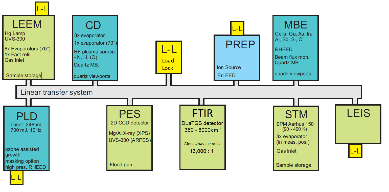
Fig. 1: UHV Cluster Layout.
1. The molecular beam epitaxy preparation chamber (UHV-MBE)
- Deposition of III-V materials (Ga, In, Al, As, Sb; C, Si for doping) for epitaxial growth of semiconductor thin films and heterostructures (GaAs, InAs) and nanowires.
- Liquid nitrogen chilled, very clean process chamber minimizing contamination by other processes. Quartz microbalance and beam flux monitor for calibration of deposition rates.
- Equipment: RHEED system for real-time layer growth measurement, sample heating by electron beam heater up to 800°C (1200 °C for short time), pyrometer for temperature measurement. Quartz viewports for attaching ellipsometer.
- Note that there is no possibility to insert your evaporator.
2. The multi-purpose chamber for the preparation of surfaces and thin layers (UHV-Deposition)
- Preparation of samples and possibility of deposition of materials by thermal evaporation (e.g. metals, organics). Quartz microbalance for calibration of deposition rates.
- Possibility to mount custom components like effusion cells.
- The chamber is equipped with an RF plasma source for atomic nitrogen and oxygen.
- Equipment: sample heating by electron beam heater up to 1400 °C, sample cooling by L-N2, pyrometer for temperature measurement.
3. The pulsed laser deposition preparation chamber (UHV-PLD)
- Preparation of oxide-based layers – possibility of epitaxial growth of oxide heterostructures
- Ozone-assisted growth, masking option.
- Equipment: UV-laser 248nm 700mJ 10Hz, high-precision RHEED, sample heating by IR laser, independent load-lock.
4. The chamber for sample heating and sputtering
(UHV-Preparation)
- Cleaning and basic preparation of the surfaces of the substrates.
- Ion sputtering (Ar ions) and annealing (e-beam heating, direct current heating – for clean Si only).
- The possibility to build-in custom components (no evaporation allowed).
- Equipment: sample heating by electron beam heater up to 1400 °C, pyrometer for temperature measurement (shared with other chambers), independent load-lock.
5. The chamber for STM and AFM scanning probe techniques
(UHV-SPM)
- Scanning Tunneling Microscopy (STM) and Atomic Force Microscopy (AFM) - achieving an atomic resolution on metal, semiconductor, and oxide surfaces.
- SPM Aarhus 150 working in range 90 – 400 K.
- Modification of the samples possible in-situ in the microscope (up to 3 evaporators). Gas inlet system.
- Equipment: Ion sources for sample and tip sputtering, electron beam heater (e-beam up to 800 °C, direct current up to 1250 °C), sample storage.
6. The chamber for photoelectron spectroscopy analysis
(UHV-XPS)
- X-ray photoelectron spectroscopy (XPS), angle-resolved XPS, ultraviolet photoelectron spectroscopy (UPS), angle-resolved photoelectron spectroscopy (ARPES).
- Analysis of the chemical composition (macroscopic area) including bonding environment of the tracked elements. Mapping of the valence band structure.
- Equipment: Mg/Al X-ray source, He UV source (He I, He II), 2D CCD detector, flood gun, high-precision computer-controlled 5-axis manipulator, sample heating by electron beam heater up to 800 °C (1200 °C for short time), sample cooling by L-He.
7. The low-energy electron microscopy system (UHV-LEEM)
- Photoelectron/Low Energy Electron Microscopy with a resolution down to 10 and 5 nanometers, respectively.
- Real-time imaging of surface processes (kinetics of the growth of layers and reactions) combined with micro-diffraction and UV spectroscopic modes for surface lattice and the electronic structure determination, respectively.
- Possibility to measure up to sample temperature of 1500 K, during evaporation or gas dosing.
- Equipment: Hg lamp, He UV source (He-I, He-II), cold-cathode electron gun (15keV), electron beam heater. Independent preparation chamber (ion source) and load-lock with sample storage.
8. The low-energy ion scattering spectroscopy system (UHV-LEIS)
- Analysis of the elemental composition of a top-most surface layer via energy spectroscopy of back-scattered noble gas ions.
- Equipment: He, Ne, Ar ion sources, full azimuth acceptance analyzer for enhanced sensitivity, atomic oxygen source for sample cleaning, independent load-lock chamber.
9. Fourier-transform infrared spectroscopy (UHV-FTIR)
- DLaTGS detector, spectral range 350 – 8 000 cm-1
- Min. 65 spectra per second
- 10 000:1 Peak-to-peak signal-to-noise ratio
- Allows for PM-IRRAS measurements


 +420 54114 9207
  nano@ceitec.vutbr.cz

