
CEITEC Nano Research Infrastructure
Brno University of Technology
Purkyňova 123, 612 00 Brno
Czech Republic
 +420 54114 9207
 nano@ceitec.vutbr.cz
Consumables | wh | ShP | i | feedback


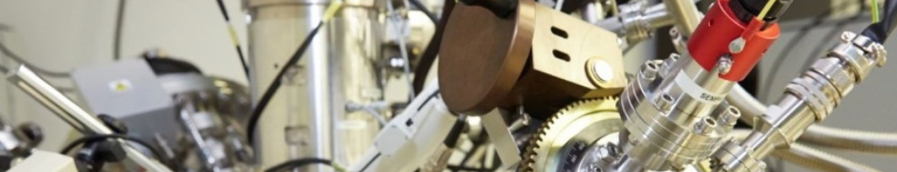
Micro & Nano X-ray CT laboratory


X-ray computed tomography is an advanced imaging technique capable of non-destructive visualization and analysis of objects. It allows scanning of the inner structure of three dimensional objects with high spatial resolution without damaging the object. Complete information about the inner structure of the object in its entire volume may be acquired from the wide range of materials, which is suitable for the detection of the shape of both internal and external structures, inhomogeneities, voids and porosities of the material.
Laboratory of X-ray micro and nano computed tomography is equipped with state-of-the art laboratory CT devices:
GE phoenix v|tome|x L240 (microCT)
|
Rigaku Nano3DX (nanoCT)
|
Computed tomography data is processed by using software: VGStudio MAX 3.1 (Volume Graphics, Germany), Avizo 9.5.0 (ThermoFisher Scientific, USA), MATLAB R2017a (MathWorks, USA), GOM inspect 2016, MAVI 1.4.1. (Fraunhofer Institute for Industrial Mathematics ITWM, Germany), 3D PDF maker Standalone.
For more information please visit our CTlab website or contact Dr. Tomáš Zikmund (tomas.zikmund@ceitec.vutbr.cz).